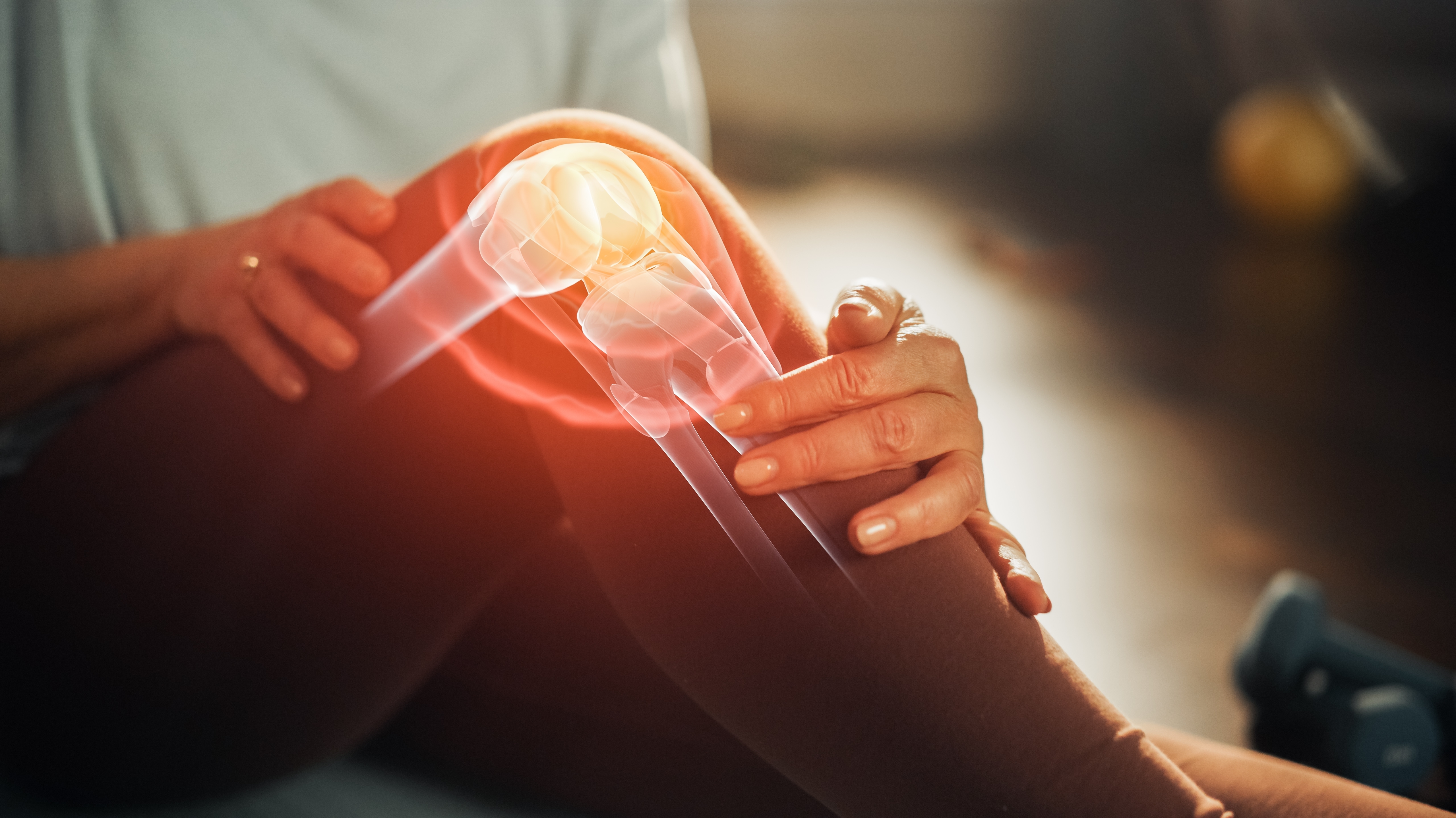
Your anterior cruciate ligament (ACL) is located in the middle of the knee joint, connecting the shin bone (tibia) to the thigh-bone (femur). It crosses over diagonally with the Posterior Cruciate Ligament (PCL) to control the backward and forward movement of the knee at the joint. It is one of four strong stabilising ligaments of the knee, along with the PCL and the medial and lateral collateral ligaments. The ACL works to stop the tibia from sliding forwards in front of the femur.
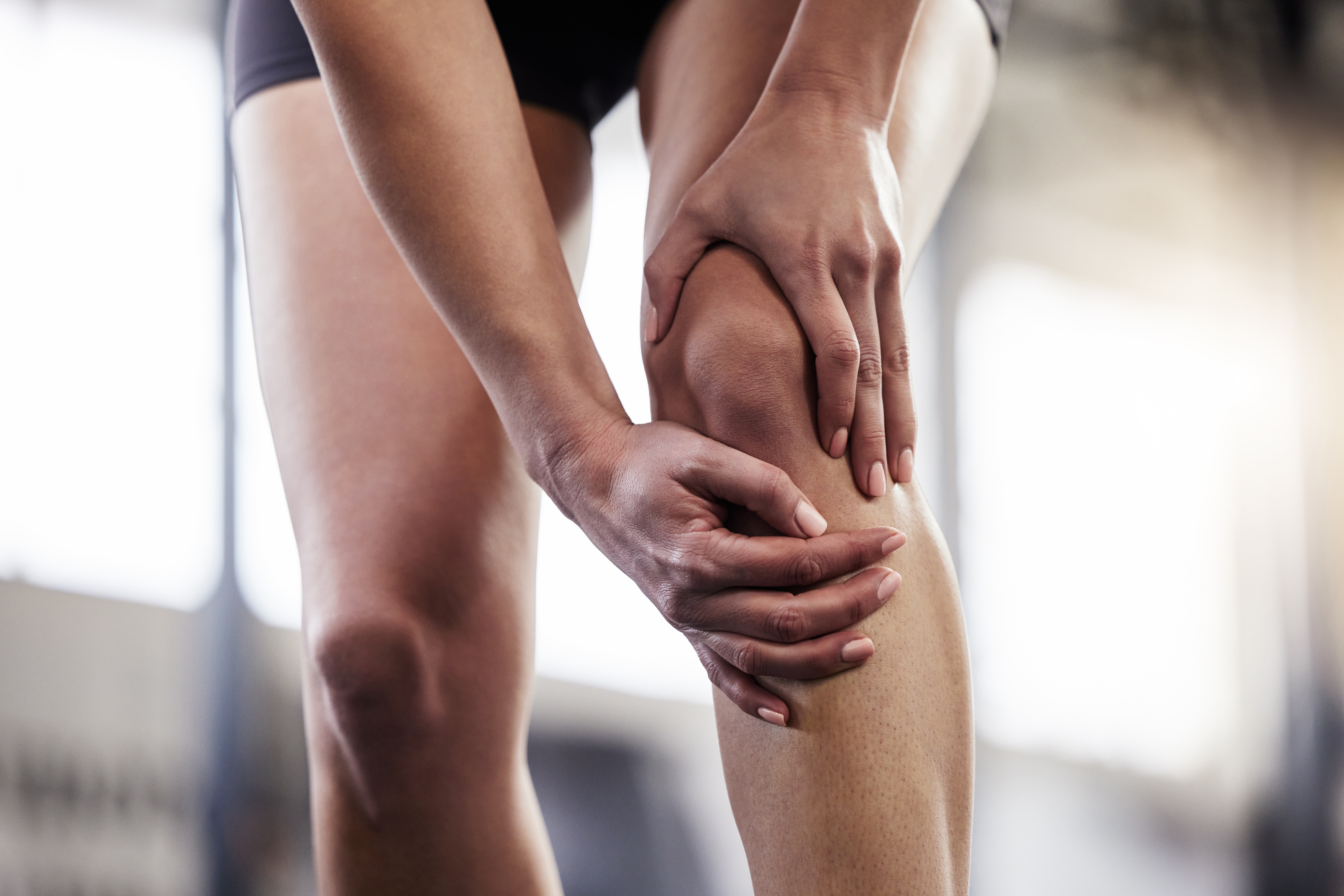
Injury to the ACL is a common knee injury, and often occurs alongside injury to other knee structures, such as the cartilage and the Collateral Ligaments. The different levels of severity of injury include a ligament sprain, a partial tear, or a complete rupture. ACL injuries are often sustained by athletes involved in high-impact sports that involve sudden changes in direction, such as basketball or soccer. ACL injuries are more common in females, which may be attributed to by muscle strength, control and conditioning. Typically, ACL injuries are caused by:
Depending on the severity of your injury, symptoms may include:
When the injury first occurs, it’s important to stop physical activity and avoid walking on the affected knee where possible. Following the PRICE principles (protection, rest, ice, compression and elevation) can help reduce the initial pain and swelling. If you have had a rupture or significant tear to your ACL, you may require surgery. It’s important that you have a diagnosis to confirm the extent of your injury, which may involve having an ultrasound, x-ray or MRI imaging. Your podiatrist can help you gradually rebuild the strength in your knee and surrounding muscles, and improve the range of motion in the joint, which will likely be restricted following your injury. Your podiatrist will make recommendations based on the cause of your injury to both help your recovery and reduce the risk of re-injury in the future. This may look at the use of Orthotics to control motion at the feet and legs, assessing the stability of your Footwear, teaching you how to strap your knee or recommending a Brace, and physical therapy.
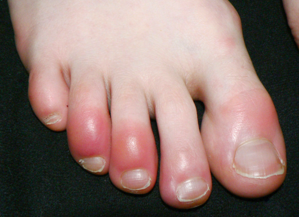
Cold, itchy, swollen toes this winter? You could be dealing with chilblains. This common condition affects the small blood vessels in your skin and can cause redness, pain, and irritation, particularly on the toes. Learn what causes chilblains, how to prevent them, and when it's time to seek professional advice.
There’s something special about a chilling morning. The quiet air, a warm cup of coffee, and a slow start before the
day begins. But for many people, that peaceful moment is interrupted by a sharp reminder the second their feet hit the floor.
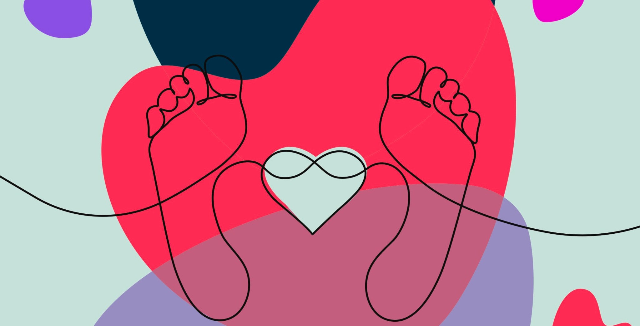
Valentine’s Day is about love, appreciation, and thoughtful gestures but let’s be honest, most gifts don’t last longer than a week. This year, choose something different. Choose a gift that offers comfort, confidence, and real care: a Medical Pedicure.
Keeping your family on their feet and helping them to walk, run, play and exceed their goals is why we love getting up in the morning.
Ground Floor, One Health Building
122 Remuera Rd, Remuera
Auckland 1050, New Zealand
| MON - FRI | 7:30am – 6:30pm |
| SAT | 8:30am – 4:30pm |
| SUN | Some availability |
Make an Appointment
Online Schedule
Our virtual receptionist is available 24/7 to help with general questions, booking requests, and clinic information, even when our team is busy, or it's after hours.
Whether you're calling us or using our website, you'll get fast assistance any time of day. And if your query needs a personal touch, a member of our team will follow up as soon as possible.
If you’d like to see a podiatrist who speaks your preferred language, just give us a call and we’ll help you book.